Home >> Satellite 2 (Department of Applied Chemistry, Chemical Engineering and Biomolecular Engineering)
Instruments of Satellite 2
| NMR1 | NMR2 |
| ESR | FT-IR |
| Fluorescence spectrometer | ICP-AES |
| Atomic absorption spectrometer | XPS |
| EPMA | FE-STEM |
| FE-SEM | Tabletop SEM |
| Mass spectrometer for proteome analysis | SC-XRD |
NMR (Nuclear Magnetic Resonance)
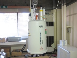
| Model (400MHz) | Bruker, AV400 (Liquid only) 1H Resonance frequency: 400MHz Probe: 5mmBBFO probe Measurable nuclides: 1H, 13C, 19F, 31P ※Please contact us for other nuclides. Measurement mode: 1-dimensional, 2-dimensional, Temperature variable |
|---|---|
| Applications | By using nuclear magnetic resonance phenomenon, it is possible to measure the chemical structure of a compound. |
| Person in charge | Kayamori, Arakawa, Furuuchi, Saito |
| Notes | Those who wish to use the equipment, please contact the person in charge of the equipment. This equipment is entrusted with maintenance and operation from "Department of Applied Chemistry, Chemical Engineering and Biomolecular Engineering" to "Instrumental Analysis Group". |
NMR (Nuclear Magnetic Resonance)

| Model (600MHz) | JEOL, ECZL-600 1H Resonance frequency: 600MHz Probe: For liquids 5mmHFX probe,For solids 3.2mmAutoMAS probe Measurable nuclides: 1H, 13C, 19F, 31P ※Please contact us for other nuclides. Measurement mode: 1-dimensional, 2-dimensional, Solids Measurement,Temperature variable |
|---|---|
| Option | Autosampler (up to 30 samples) |
| Applications | By using nuclear magnetic resonance phenomenon, it is possible to measure the chemical structure of a compound. |
| Person in charge | Kayamori, Arakawa, Furuuchi, Saito |
| Notes | Those who wish to use the equipment, please contact the person in charge of the equipment. This equipment is entrusted with maintenance and operation from "Department of Applied Chemistry, Chemical Engineering and Biomolecular Engineering" to "Instrumental Analysis Group". |
ESR (Electron Spin Resonance)
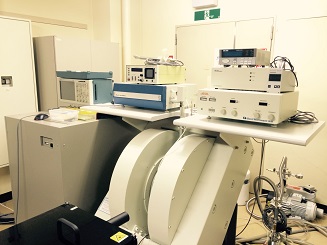
| Model | JEOL, X330 |
|---|---|
| Microwave frequencies | 8.75 - 9.65GHz (X-band) 34 - 36GHz (Q-band) |
| Maximum field strength | 1.4 T |
| Maximum microwave power | 200mW |
| Applications | Obtain information on unpaired electrons in a substance by using the electron spin resonance phenomenon. |
| Person in charge | Kayamori, Arakawa, Furuuchi, Saito |
| Notes | Those who wish to use the equipment, please contact the person in charge of the equipment. This equipment is entrusted with maintenance and operation from "Department of Applied Chemistry, Chemical Engineering and Biomolecular Engineering" to "Instrumental Analysis Group". |
FT-IR (Fourier transform infrared spectrometer)
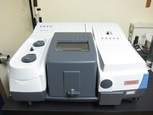
| Model | ThermoFisher, Nicolet6700 |
|---|---|
| Wavenumber range | 7400 - 350cm-1 |
| Resolution | 0.09cm-1 |
| Methods | Transmission method, Diffuse reflection method, ATR method |
| Applications | Chemical structure analysis of various samples. |
| Person in charge | Kayamori, Arakawa, Furuuchi, Saito |
| Notes | Those who wish to use the equipment, please contact the person in charge of the equipment. This equipment is entrusted with maintenance and operation from "Department of Applied Chemistry, Chemical Engineering and Biomolecular Engineering" to "Instrumental Analysis Group". |
Fluorescence spectrometer
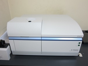
| Model | Hitachi High-Technologies, F-7000 |
|---|---|
| Sensitivity | Over S/N 250 |
| Light source | 150W Xe lamp |
| Measurement wavelength range | 200 - 900nm, and zero-order light |
| Resolution | 1.0nm |
| Minimum sample volume | 0.6mL (in use of standard 10 mm rectangular cell) |
| Modes | fluorescence, Phosphorescence, luminescence, 3-dimensional measurement |
| Optional unit | Constant temperature unit Low temperature (liq. N2 temp.)unit Solid unit |
| Applications | Perform qualitative and quantitative analysis using fluorescence emitted when the sample in the excited state returns to the ground state. |
| Person in charge | Kayamori, Arakawa, Furuuchi, Saito |
| Notes | Those who wish to use the equipment, please contact the person in charge of the equipment. This equipment is entrusted with maintenance and operation from "Department of Applied Chemistry, Chemical Engineering and Biomolecular Engineering" to "Instrumental Analysis Group". |
ICP-AES (Inductively Coupled Plasma Atomic Emission Spectrometer)
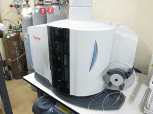
| Model | ThermoFisher, iCAP6500 |
|---|---|
| Spectrometer | Simultaneous echelle type |
| Wavelength range | 166.25 - 847nm |
| Spectral bandpass | 0.007nm at 200nm |
| RF source | 0.75 - 1.6kW (Duo restricted to 1.35 kW) |
| Applications | The sample is atomized and excited by inductively coupled plasma, and the qualitative and quantitative determination of elements is performed from the emission spectrum obtained when returning to the ground state. |
| Person in charge | Kayamori, Arakawa, Furuuchi, Saito |
| Notes | Those who wish to use the equipment, please contact the person in charge of the equipment. This equipment is entrusted with maintenance and operation from "Department of Applied Chemistry, Chemical Engineering and Biomolecular Engineering" to "Instrumental Analysis Group". |
Atomic absorption spectrometer
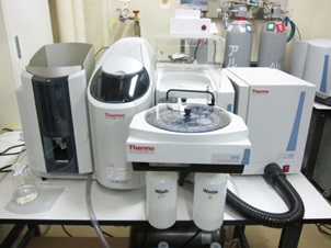
| Model | ThermoFisher, iCE3500 |
|---|---|
| Wavelength range | 180 - 900nm |
| Resolution | 0.5nm/mm |
| Applications | The concentration of the target element is determined from the amount absorbed when atoms in the ground state absorb light of a specific wavelength and excite it. |
| Person in charge | Kayamori, Arakawa, Furuuchi, Saito |
| Notes | Those who wish to use the equipment, please contact the person in charge of the equipment. This equipment is entrusted with maintenance and operation from "Department of Applied Chemistry, Chemical Engineering and Biomolecular Engineering" to "Instrumental Analysis Group". |
XPS (X-ray Photoelectron Spectrometer)
| Model | Shimadzu, AXIS-ULTRA |
|---|---|
| X-ray source | Achromatic Al/Mg Kα |
| Minimum spot size | 15µm⌀ or less |
| Sample size | 100mm×40mm×20mm |
| Applications | The energy of photoelectrons emitted when the sample is irradiated with X-rays is measured to analyze constituent elements and chemical bonding state of the solid surface. |
| Person in charge | Kayamori, Arakawa, Furuuchi, Saito |
| Notes | Those who wish to use the equipment, please contact the person in charge of the equipment. This equipment is entrusted with maintenance and operation from "Department of Applied Chemistry, Chemical Engineering and Biomolecular Engineering" to "Instrumental Analysis Group". |
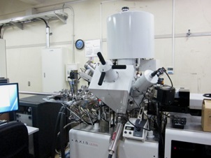
EPMA (Electron Probe Micro Analyzer)
| Model | Shimadzu, EPMA-1720HT |
|---|---|
| Electron source | CeB6 cathode |
| X-Ray Spectrometers | 5 channels (10 analyzing crystals) |
| Sample size | ⌀25mm×20mmh |
| Analyte elements | B - U |
| Accesory devices | EDS (analyte elements: B - Am) |
| Applications | From the wavelength of the characteristic X-ray generated when the sample is irradiated with the electron beam, the constituent elements, distribution state, concentration, etc. of the solid surface are analyzed. |
| Person in charge | Kayamori, Arakawa, Furuuchi, Saito |
| Notes | Those who wish to use the equipment, please contact the person in charge of the equipment. This equipment is entrusted with maintenance and operation from "Department of Applied Chemistry, Chemical Engineering and Biomolecular Engineering" to "Instrumental Analysis Group". |
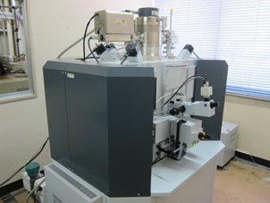
FE-STEM (Field Emission Scanning Transmission Electron Microscope)
| Model | Hitachi High-Technologies, HD-2700 |
|---|---|
| Accelerating voltage | 200kV, 120kV, 80kV |
| Image resolution | 0.204nm (magnification: ×4000k, HR mode) |
| Magnification | 100 to 10,000k× |
| Applications | An electron beam is transmitted through the specimen to observe the nano-level microstructure. |
| Person in charge | Kayamori, Arakawa, Furuuchi, Saito |
| Notes | Those who wish to use the equipment, please contact the person in charge of the equipment. This equipment is entrusted with maintenance and operation from "Department of Applied Chemistry, Chemical Engineering and Biomolecular Engineering" to "Instrumental Analysis Group". |
FE-SEM (Field Emission Scanning Electron Microscope)
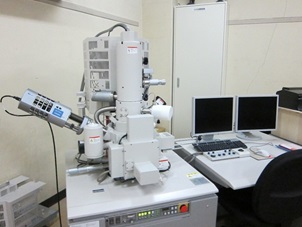
| Model | Hitachi High-Technologies, S-4800 |
|---|---|
| Accelerating voltage | 0.5 - 30kV |
| Image resolution | 1.0nm (15kV, WD: 4mm) |
| Magnification | 100 to 800,000× |
| Applications | Secondary electrons emitted when the sample is irradiated with an electron beam are detected, and the shape of the fine region is observed. |
| Person in charge | Kayamori, Arakawa, Furuuchi, Saito |
| Notes | Those who wish to use the equipment, please contact the person in charge of the equipment. This equipment is entrusted with maintenance and operation from "Department of Applied Chemistry, Chemical Engineering and Biomolecular Engineering" to "Instrumental Analysis Group". |
Tabletop SEM (Tabletop Microscope)
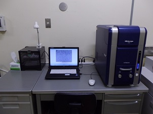
| Model | Hitachi High-Technologies, TM3000 |
|---|---|
| Accelerating voltage | 5kV, 15kV |
| Magnification | 15 to 30,000× |
| Detector | Backscattered electron detector |
| Observation mode | Standard mode, Charge-up reduction mode |
| Applications | Secondary electrons emitted when the sample is irradiated with an electron beam are detected, and the shape of the fine region is observed. |
| Person in charge | Kayamori, Arakawa, Furuuchi, Saito |
| Notes | Those who wish to use the equipment, please contact the person in charge of the equipment. This equipment is entrusted with maintenance and operation from "Department of Applied Chemistry, Chemical Engineering and Biomolecular Engineering" to "Instrumental Analysis Group". |
Mass spectrometer for proteome analysis
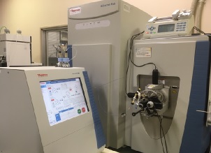
| Model | Thermo Fisher Scientific, Q Exactive Plus |
|---|---|
| MS type | LC-MS/MS |
| LC part | Easy n-LC1000 |
| Mass spectrometer part | Quadrupole / Orbitrap hybrid |
| Ionization | ESI |
| Maximum resolution | 140,000 (m/z 200) |
| Mass range | m/z 50~6000 |
| Mass accuracy | Less than 3ppm (external standard), Less than 1ppm (internal standard) |
| Applications | Proteomic analysis of biological samples. |
| Person in charge | Kayamori, Arakawa, Furuuchi, Saito |
| Notes | Those who wish to use the equipment, please contact the person in charge of the equipment. This equipment is entrusted with maintenance and operation from "Department of Applied Chemistry, Chemical Engineering and Biomolecular Engineering" to "Instrumental Analysis Group". |
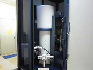
SC-XRD
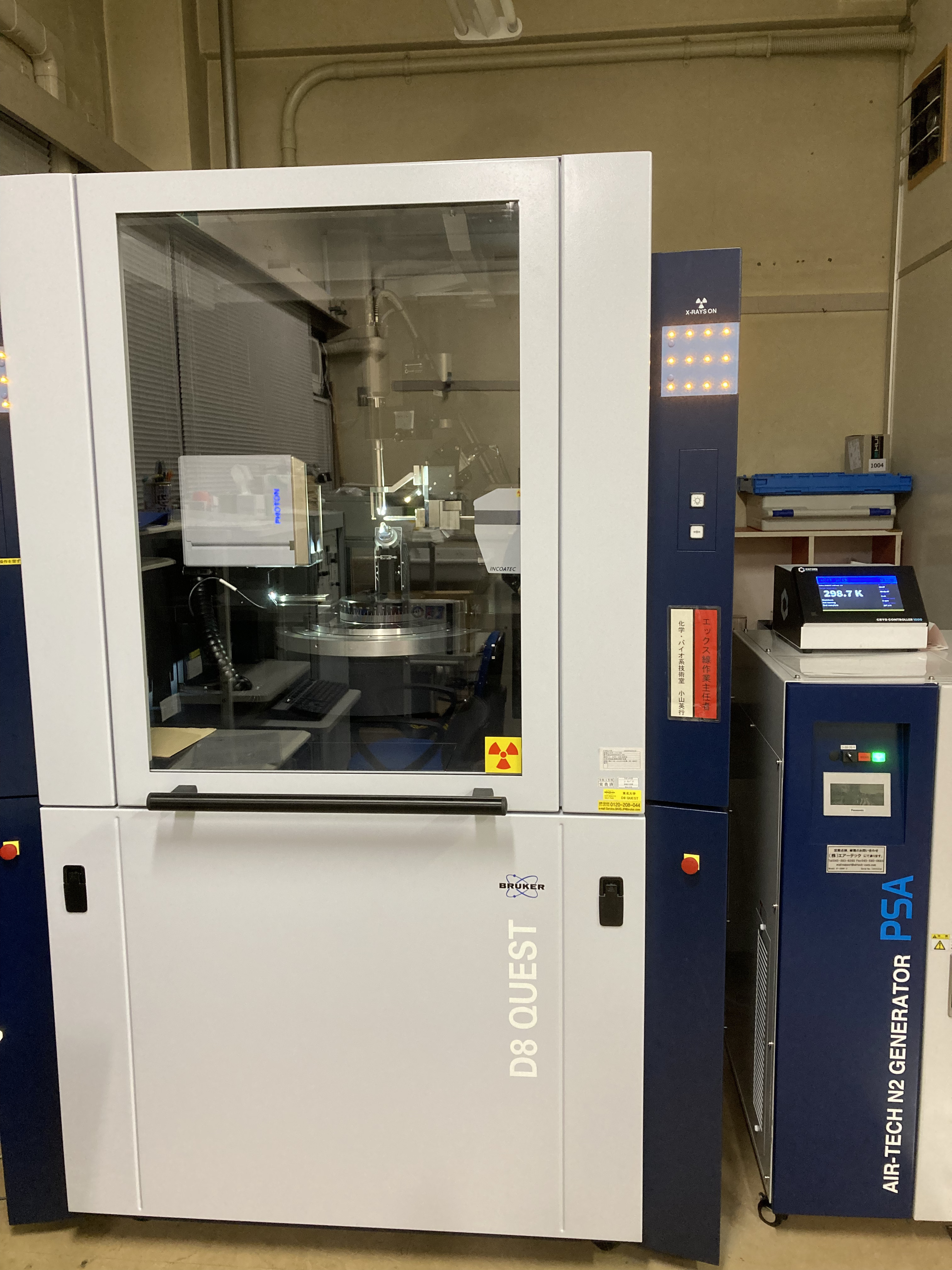
| Model | Bruker D8 QUEST |
|---|---|
| X-Ray source | Mo target, μ focus type |
| Detector | Two-dimensional semiconductor detector(100×70mm) |
| Option | Temperature variable(90-400K) |
| Sample crystal size | < 0.2mm square |
| Applications | Analysis of the molecular structure of crystals. |
| Person in charge | Kayamori, Arakawa, Furuuchi, Saito |
| Notes | Those who wish to use the equipment, please contact the person in charge of the equipment. This equipment is entrusted with maintenance and operation from "Department of Applied Chemistry, Chemical Engineering and Biomolecular Engineering" to "Instrumental Analysis Group". |